The concept that chronic skin inflammation may be the source of skin inflammaging is expanding and so natural ingredients that can address sources of skin inflammation are desired. Chronic skin inflammation driven by activation of caspase-1 via skin cell NLRP (Nod-Like Receptor Protein) inflammasome activation is a strategic target for new skin care technologies.
Over two decades ago, two important concepts of aging seemed to emerge almost simultaneously. The first was the idea that aging, including skin aging, may be driven by processes of chronic inflammation. This idea spawned the term “inflamm-aging” which was coined by Professor Claudia Franceschi at the University of Bologna1. The second groundbreaking event was the discovery by Professor Jurg Tschopp and colleagues at the University of Lausanne in Switzerland of the Nod-Like Receptor Proteins (NLRP)2. These unique inflammatory proteins are critical for unlocking the mechanisms by which the body’s innate immune response occurs, initiating processes of inflammation in the body. It soon became apparent that a link exists between the NLRP inflammasomes and body and skin inflammaging and numerous papers appeared that linked these two concepts together3-8. In 2019, Gruber et al, published work that outlined the benefits of various botanical blends to address the NLRP Inflammasome-activated release of caspase-1 from Normal Human Epidermal Keratinocytes [NHEK] and Reconstructed Human Epidermis [RHE] models stimulated by UVB and ATP9,10. From this work, a series of three unique botanical blends, known by the trade name “Strataphix”, were launched in 2020. “Strataphix Poly” [INCI Name: Water (and) Glycerin (and) Laminaria Digitata Extract (and) Opuntia Ficus Indica Stem Extract (and) Galactoarabinan (and) Schizophyllan, polysaccharide blend] emerged as a unique topical ingredient that demonstrated advantages at controlling skin irritation and also was able to control the significant dehydrating impacts of retinol on the skin11,12. Since this initial launch, the ingredient has begun to appear in various commercial products principally in the United States. Part of the difficulty with launching the ingredient in Europe and Asia was the original product was preserved with phenoxyethanol which is now frowned upon in the EU and Asia. After working on the formulation, Vantage has now developed a preservative-free version of the polysaccharide-based blend called “Strataphix Poly-PF”. This new form of the polysaccharide blend has been shown to be microbially stable by using 1,2-hexanediol and pentylene glycol as bacteria controlling ingredients.Vantage continues to examine the benefits of all three botanical blends with additional testing. However, considerable focus has been on examining the continuing benefits of the polysaccharide blend through in vitro assays examining the blend’s influence on skin irritation from Cutibacterium acnes and more recently by clinical examination of the ingredient for its ability to improve highly melanated skin of people of color.13,14 This paper will examine these latest studies that continue to support the profound benefits of this unique blend of potent polysaccharides on skin.
Materials and Methods
In vitro Cutibacterium acnes inflammation testing of polysaccharide blend
The methods employed to examine the effects of the polysaccharide blend on RHE pre-treated with Cutibacterium acnes, salicylic acid and the polysaccharide blend have been outlined in detail in an earlier publication13. Briefly, RHE (“Epiderm”, Mattek) was treated as shown in figure 1.The resulting tissues were examined for expression of caspase-1 and IL-1β using standard ELIAS protein assays. The studies were run in triplicate and the results were analyzed statistically using the Student Paired T-Test in Excel.
Clinical examination of polysaccharide blend on highly melanated individuals of color
The methods employed in the following study were outlined in an earlier NYSCC presentation14. Briefly, a clinical study was initiated on a panel of 38 individuals with Fitzpatrick skin types of III-VI following appropriate global testing protocols. The study progressed for 56-days with the individuals broken into two groups of 19 participants: Placebo formulation without polysaccharide blend and Active formulation with 1.0% of the polysaccharide blend which the participants applied 2X/Day around the face. The participants were tested on Day 28 and Day 56 for skin moisturization and were given a subjective evaluation on Day 56 to assess their feelings about the formulations. Photographs of all the participants were taken on Day 28 and 56 using the Visia CR camera system. Statistical analysis on the skin moisturization measurements was done using the Student Paired T-Test.
Results and Discussion
The results of the examination of the treatment of 1.0 and 3.0% of the polysaccharide blend on Epiderm tissues pre-treated for 48-hours with Cutibacterium acnes and then for 24-hours with either a formulation containing 1.0 or 2.0% salicylic acid or a formulation containing 1.0% salicylic acid and 2.0% salicylic acid with 1.0% or 3.0% of the polysaccharide blend, respectively, is shown in figure 2. The data from figure 2 indicates that the treatment of the tissues with Cutibacterium acnes for 48-hours followed by treatment of the tissues with 1.0% or 2.0% salicylic acid results in an increased expression of active caspase-1 measured using the same ELISA test methods reported in the earlier work by Gruber et al, above the results of the negative controls (data not shown)13. Caspase-1 is a potent protease enzyme that is released principally via the NLRP3 inflammasome activation in dermal and epidermal skin cells. Release of active caspase-1 initiates the downstream activation of numerous inflammatory cytokines, principally IL-1β and IL-18. The data for examination of the release of active IL-1β is shown in figure 3.Interluken-1β (IL-1β) exists within the skin cells in an inactive form called proIL-1β2. Release of caspase-1 allows the inactive form of IL-1β to become activated which then begins processes of downstream irritation within the skin, driven by the skin’s innate immune response that does not require previous biological imprinting of the irritating external targets, Cutibacterium acnes and salicylic acid, on the skin.From the data in figure 3, again it can be seen the topical treatment of the previously Cutibacterium acnes-irritated tissues with the treatment with 1.0 and 2.0% salicylic acid further elicits the release of active IL-1β above that of the negative control (data not shown). Treatment of the tissues with 1.0 and 2.0% salicylic acid in addition to 1.0 and 3.0% of the polysaccharide blend, respectively, results in a statistically significant reduction in the release of the IL-1β within the treated tissues compared against the salicylic acid treated tissues indicating that the polysaccharide can help mitigate the irritating effects of the Cutibacterium acnes and salicylic acid on the tissues.The results of the skin hydration measurements taken at Days 28 and 56 for each group of 19 individuals treated in the clinical study are shown in figure 4.The results of the moisturization measurements demonstrate and continue to support the moisturizing benefits of the polysaccharide blend noted in earlier work11,12. Compared against the Placebo cream without the polysaccharide blend, the formulation containing 1.0% of the polysaccharide blend showed statistically significant skin moisturization on individuals with darker skin (Fitzpatrick Type III-VI). When quizzed at the end of the study with a subjective evaluation, the individuals using the formulation containing the polysaccharide blend showed a preference over the formulation without the polysaccharide blend, figure 5.Figure 6 shows the image of one of the individual participants from the study showing improvements in skin appearance on Day 28 and Day 56 in the study.
Conclusion
The data presented in this paper was the culmination of several expanded studies which more deeply examined the mechanisms by which the unique polysaccharide blend can reduce inflammasome-mediated release of key inflammatory molecules in irritated skin. The data is further supported by clinical studies on individuals with highly melanated skin demonstrating that the polysaccharide blend can help improve the appearance and moisturization of the skin within 28 Days. Importantly, the ingredient appears to work well in combination with salicylic acid, a popular skin ingredient that has demonstrated benefits in troubled skin but may carry some inflammatory propensities itself.Acknowledgments: The authors appreciate the help of VitroScreen in running the in vitro Cutibacterium acnes studies and CompLife for performing the clinical studies on individuals with highly melanated skin.
References:
1 Franceschi C, et al., Inflamm-aging. An evolutionary perspective on immunosenescence. Ann N Y Acad Sci. 2000 Jun:908:244-54. doi: 10.1111/j.1749-6632.2000.tb06651.x.
2 Martinon F, et al., The Inflammasome: A Molecular Platform Triggering Activation of Inflammatory Caspases and Processing of proIL-β. Mol Cell. 2002 Aug;10(2):417-26. doi: 10.1016/s1097-2765(02)00599-3.
3 Latz E et al., NLRP3 inflammasome activation in inflammaging. Semin Immunol. 2018 Dec:40:61-73. doi: 10.1016/j.smim.2018.09.001.
4 Marin-Aguilar F et al., Aging and the inflammasomes. Exp Suppl. 2018:108:303-320. doi: 10.1007/978-3-319-89390-7_13.
5 Cordero MD et al., Inflamm-ageing or inflammasom-ageing as independent events. Aging (Albany NY). 2020 Sep 28;12(18):17759-17760. doi: 10.18632/aging.104108.
6 Cyr B et al., The Role of Non-canonical and Canonical Inflammasomes in Inflammaging. Front Mol Neurosci. 2022 Feb 9:15:774014. doi: 10.3389/fnmol.2022.774014.
7 Mejias NH et al., Contribution of the inflammasome to inflammaging. J Inflamm (Lond). 2018 Nov 16:15:23. doi: 10.1186/s12950-018-0198-3.
8 Meyers AK et al., The NLRP3 Inflammasome: Metabolic Regulation and Contribution to Inflammaging. Cells 2020: 9(8):1808; doi.org/10.3390/cells9081808.
9 Gruber JV et al., In vitro expression of NLRP inflammasome-induced active Caspase-1 expression in normal human epidermal keratinocytes (NHEK) by various exogenous threats and subsequent inhibition by naturally derived ingredient blends. J Inflamm Res. 2019 Aug 26:12:219-230. doi: 10.2147/JIR.S215776.
10 Gruber JV, et al., Topical Botanical Complex Formulations Designed to Inhibit the Impact of Free Radicals on Skin Aging Processes. Eurocosmetics. 2020 Sept:9:10-13.
11 Gruber JV et al., NLRP inflammasomes and induced skin inflammation, barrier recovery and extended skin hydration. Int J Cosmet Sci. 2020 Feb;42(1):68-78. doi: 10.1111/ics.12588.
12 Gruber JV et al., Retinol Has a Skin Dehydrating Effect That Can Be Improved by a Mixture of Water-Soluble Polysaccharides. Cosmetics 2020: 7(4):80. doi.org/10.3390/cosmetics7040080.
13 Gruber JV et al., A mixture of water-soluble polysaccharides reduces caspase-1 and IL-1β inflammatory responses by Cutibacterium acnes in vitro in Reconstructed Human Epidermis (RHE). Clin Cosmet Invest Dermatol 2024:17:2627-2633. Doi.org/10.2147/CCID.S487881.
14 STRATAPHIX® POLY: An Advanced Skin Inflammaging Treatment with Superior Clinical Moisturizing Benefits on Melanated Skin. NYSCC Melanated Skin Symposium. September 2024. Poster.
James V Gruber, Nicole Terpak, Sebastien Massard, Elva Chen, Yurah Kim, Maeva Abdoulhadi Vantage Specialty Chemicals, Warren/NJ, USA, www.vantagegrp.com
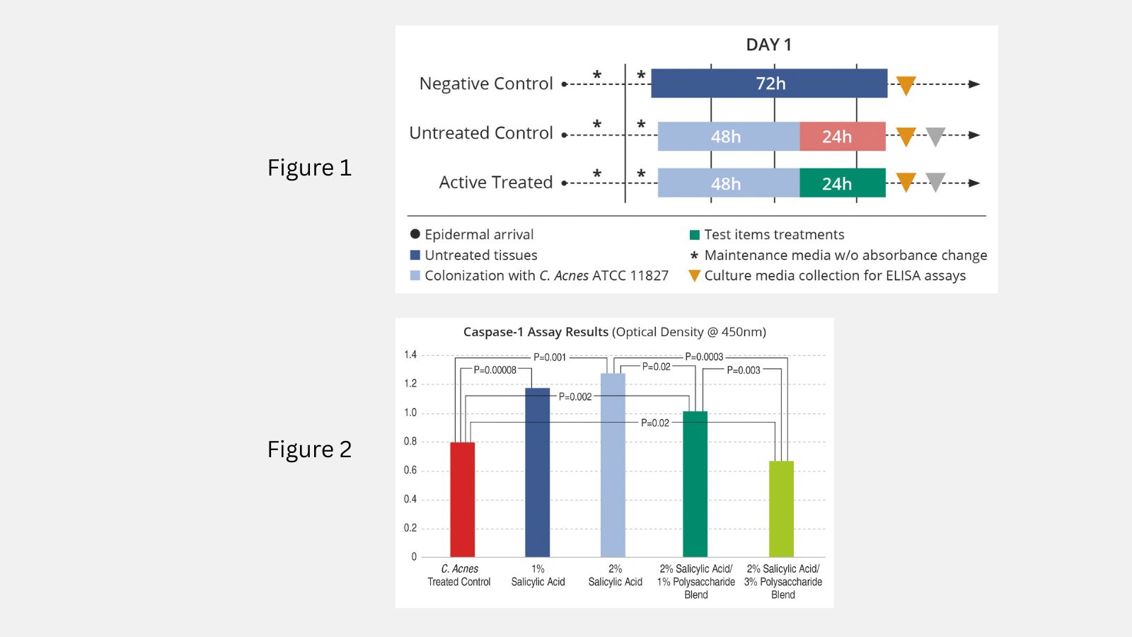
Figure 1
Schematic summary of methods of treatment of “Epiderm” tissues with Cutibacterium acnes for 72 hours, (negative control), Cutibacterim acnes and 1 or 2% salicylic acid (untreated control) or Cutibacterium acnes, 1 or 2% salicylic acid and 1% of the polysaccharide blend (active treated). Prior to treatment of the tissues with the topical ingredients, the Cutibacterium acnes was removed by rising with Phosphate Buffer Solution.
Figure 2
Results of treatment of Epiderm RHE tissue models with Cutibacterium acnes for 48-hours followed by either 1.0 or 2.0% salicylic acid alone, or the same concentrations of salicylic acid with 1.0 and 3.0% of the polysaccharide blend, respectively, examining release of active caspase-1. For clarity, the results of the treatment of each tissue with the Cutibacterium acnes alone (negative control) were excluded as all data points shown in the graph were statistically significant against the negative control. p≤0.05.
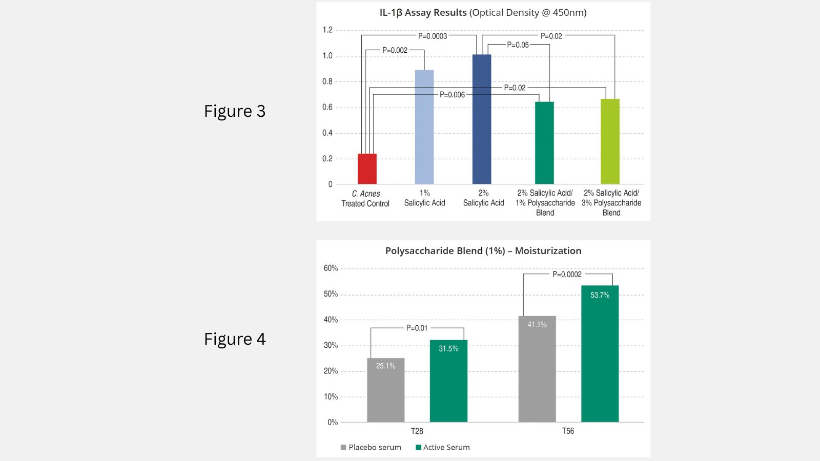
Figure 3
Results of treatment of Epiderm RHE tissue models.
Figure 4
Results of skin moisturization testing on Day 28 and Day 56. Placebo serum (grey), Active Serum (dark green), N=38, p≤0.05.
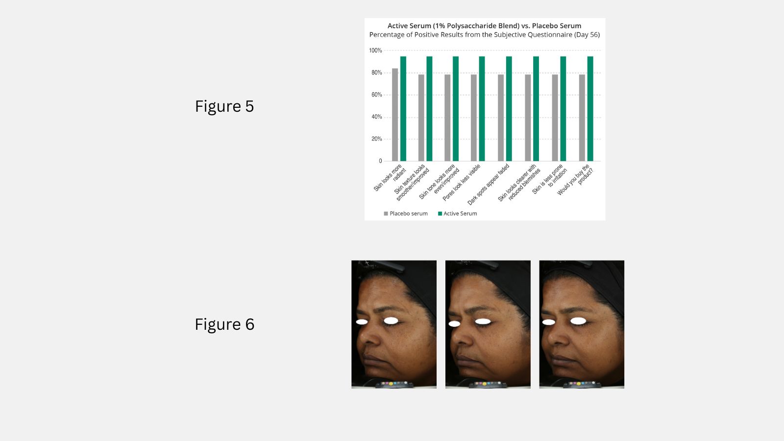
Figure 5
Subjective Questionnaire Results in Percentage on Day 56. Placebo Serum (grey), Active Serum (dark green).
Figure 6
VISIA-CR images of volunteer from study showing progression (left to right) from Baseline to Day 28 and Day 56.