
The microflora, or as we now call it more precisely microbiota, is in fact the assemblage of microorganisms which are present in a defined environment3. The human microbiota is composed of a huge diversity of bacteria, yeasts, fungi and viruses, and their total number is estimated to be 10,000 different species. Bacterial communities are extraordinarily complex, and this applies to the bacterial communities of our body as well, inhabiting all of our organs, including the gut, the mouth, the nose, the vagina, skin and even blood. These organs provide various conditions for bacterial life – such as moisture or dryness, available nutrients, pH and temperature.
For instance, typical bacterial compositions have been identified as altered lung microbiomes that are associated with cystic fibrosis, chronic obstructive pulmonary disease, or asthma. There are also associations between unbalanced intestinal tract microbiomes and individuals with cystic fibrosis and diabetes4, 5.
Our skin provides many niches with different ecological conditions (humidity, pH, available nutrients) and is also exposed to a large variety of external parameters, such as outside temperature, UV-exposure, pollution and even contact. This wide variety of skin ecosystems results in many differences in cutaneous microbial composition and abundance depending on the body area, and all of this constitutes the skin microbiome6, 7.
Elizabeth Grice and Julia Segre in 2011 did a publication on the diversity of the microbiome on the skin, our largest organ8. Microbes on our skin also communicate with our body,9 and in particular play an important role in our immune system, as explained by Julia Segre 10. As a prolific expert in the field, Richard Gallo also demonstrated the link between cutaneous microbiota and skin health11.
Probiotics are living microorganisms that are supplied to the gut (via ingestion) to rebalance an altered microbiome and modify its equilibrium towards a healthier one. The classical Bifidobacterium or Lactobacillus we can find in yoghurt, for instance, are typical probiotics. Prebiotics, on the other hand, are food ingredients that promote the growth of beneficial microorganisms in the intestines3.
“The skin’s composition is the consequence of our genetics, diet, life style and the area we live in”
Dr Bernd Becker, Consultant, MolCareConsulting
Beauty and the skin microbiome
The skin microbiome is without doubt a full player in our beauty and our well-being. Understanding the direct effects of cosmetic ingredients on the skin microbiome links the skin microbiome directly to the attainment of beauty. But how do we assess it, how do we evaluate the impact of a cosmetic ingredient or end-product on the Stratum microbium?
At companies such as Givaudan Active Beauty for example, the R&D team is experienced in both techniques – assessing the skin microbiome and evaluating the impact of ingredients on it. After all there is a particular knowhow on cutaneous microbiota sample collection, as well as proprietary genes databases that are exploited with home-made bioinformatics pipelines. Indeed, after many years spent in developing procedures and databases to explore soil and gut microbiomes, the company focused on skin metagenomics techniques and were one of the first to publish on this very specific topic13, 14.
Cosmetics and skin microflora
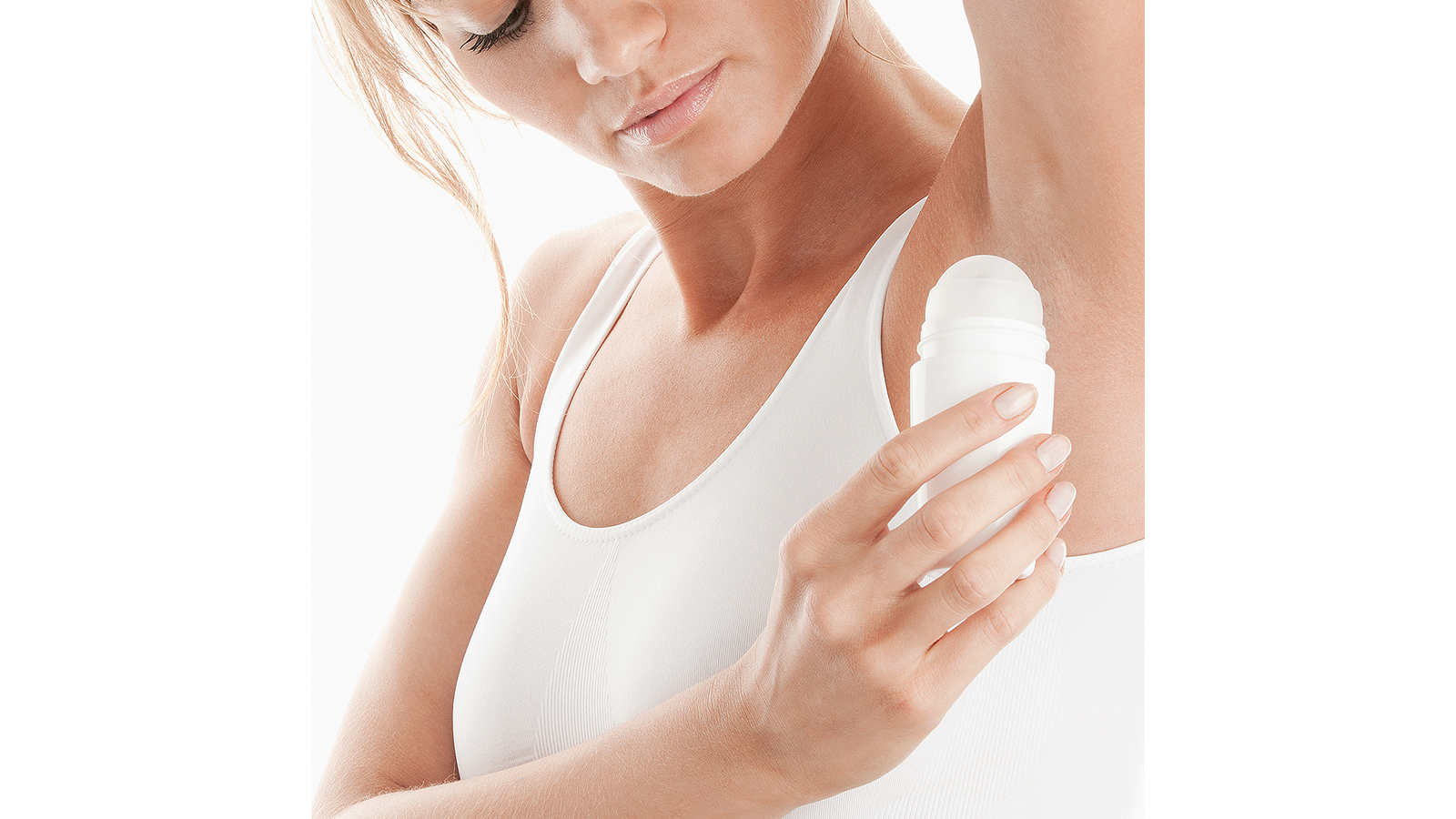
One additional role of microbes living on our skin is the production, or modulation, of body odours, in particular to produce axillary malodours by decomposing apocrine sweat (usually by Corynebacteria), or foot odours from eccrine sweat3, 12. Deodorants were thus the first cosmetic products developed to have an effect on our skin microbiota, as were new therapies to treat atopic dermatitis or rosacea, or even dandruff3.
How does acne develop?
Acne is a disease of the sebaceous glands with purulent pustules and blackheads. Many factors lead to the creation of this skin disease. During adolescence of females and males there is an increase of androgens in the blood. The increased androgens lead to an increase in sebum, which leads to greasy skin and hair. Simultaneously, the follicles are blocked by terminally differentiated dead keratinocytes, the horn cells. This induces the formation of a blackhead. Microbiota from the skin flora, particularly Propionibacterium acnes, proliferate very rapidly and provoke an inflammatory reaction of the follicle15.
The impact of the colon’s microbiota on the skin
The interaction between colon and skin is easily comprehensible15. The skin does not stop at the lips but links the skin of the face to the inside of the body through the entire digestive system. Therefore, skin and the colon mucosa are in direct and tight connection. The skin as well as the colon mucosa define the borderlines between our environment and our bodies both inside and out, protecting us from harmful impacts.
This is why the flora of the colon is of major importance. The microbiota of the colon, “the microbiome” not only decide whether we have a thick or thin body but very often about its health status. The colon’s wide-ranging effect reaches far inside the body, influencing nearly every organ. The colon behaves in a way like a serviceable, yet outgoing friend. The microbiota produces messenger molecules and hormones and in addition coaches the immune cells. Keeping the colon healthy is a major prerequisite for maintaining one’s health. A healthy colon leads not only to a good body mass index and general happiness, but also and even more to improving the skin e.g. promoting a proper complexion, fewer pustules, full hair, less allergic reactions and alleviation of skin diseases.
The skin microbiome
The compounds of the skin flora are permanently changed and regularly complemented, e.g. by shaking hands. Microbiota are regularly exchanged between humans. However, the incredibly huge number and diversity of microorganisms on our skin protects us against harmful pathogens.
The inflammatory process has a very important function in the immune system. An inflammation protects against the attempts of pathogens to enter the body. What is interesting is the fact that in advance of a disease and a strong inflammation, the bacterial diversity often decreases weeks, months or even years before the disease occurs. Harmful bacteria can spread more easily and suppress protecting microbiota. Correlations were found between neurodermitis, allergies and diabetes. It is known, that the colon’s flora during normal ageing deteriorates and turns into an impotent monoculture.
Polyphenols – the florist of the colon
Polyphenols are secondary compounds from plants responsible for the colour, taste and smell of food products. Many studies demonstrate that polyphenols effectively absorb harmful substances (free radicals) and support and increase the intrinsic protection mechanisms of the skin against sunlight (UV-radiation), delay of wrinkling and senile lentigo formation. They also lead to a radiant complexion. Due to their dichotomy, polyphenols help to improve health16 with their biotransformation of their metabolites and the modulation of colon’s microbiota.
Most of the polyphenols pass together with food through the small intestine without being absorbed. They reach the colon and have an impact on the colon’s microbiome,17 so that a reciprocal connection and interaction between polyphenols and the colon’s microbiome is created.
Skin microbes induce hair growth
Early in the life of neonatal skin, there is a short period of time during which the first steps for hair growth are started by the accumulation of regulatory T cells (Tregs) that mediate tolerance to commensal skin bacteria18. The Treg migration into the neonatal skin drives hair follicle morphogenesis19. The commensal bacteria enter the hair follicle, and together with invading Tregs, enter the new hair follicle. The hair follicle (HF) development facilitates the accumulation of Tregs in neonatal skin and organises a primary reservoir for skin commensals.
In the skin, there is a constant dialogue between the immune system and commensal bacteria. The skin is the interaction area between commensal microbes fundamental to skin physiology and immunity. Regulatory T (Treg) cells play a critical role in establishing and maintaining immune homeostasis in peripheral tissues. In the colon, commensals themselves facilitate the generation of the peripherally induced Treg cells that are critical for proper immune homeostasis.
Treg cell accumulation in neonatal skin leads to hair follicle development. However, the molecular mechanisms that allow us to tolerate the presence of skin commensals without eliciting destructive inflammation are still unknown. A defined period of neonatal life is characterised by an abrupt influx of highly activated regulatory T (Treg) cells into neonatal skin. During this period of time the host-commensal relationship in the skin relies on a unique Treg cell population that mediates tolerance to bacterial antigens18.
| Diversity |
|---|
| The human microbiota is composed of a huge diversity of bacteria, yeasts, fungi and other viruses |
The microbiome and anti-ageing
The study published in 2017 by Bing Han et al. describes the influence of the microbial composition of the microbiome on ageing20. They found that certain bacterial genes and compounds can slow the progression of tumors and the accumulation of amyloid-beta, a compound associated with Alzheimer’s disease. The study’s researchers posit that in the end, slowing down the ageing process entirely may be possible with supplements derived from gut bacteria21.
Skin microbiota–host interactions
The skin is a dynamic ecosystem inhabited by skin microbes (bacteria, archea, fungi and viruses). These microbes are fundamental to skin physiology and immunity9.
The skin, on the other hand, is also replete with diverse and unusual lipids not found elsewhere in the body22, 23. Some of these lipids, such as sapienic acid, can have antimicrobial activities, while others, such as triglycerides, can be metabolised by microbes into free fatty acids and di- and monoglycerides that can be bioactive against other microbes or stimulatory to host cells24, 25. Sanford and colleagues showed that short-chain fatty acids (SCFAs) produced by microbes colonising the skin surface lead to the inhibition of histone deacetylase (HDAC) activity. It is known from microbes that populate the intestinal tract that they produce high levels of SCFAs that inhibit HDAC activity26-28.
These findings support clinical associations between the microbial metabolome of the gut and normal immune function and provide one mechanistic explanation for how bacteria can regulate inflammation1, 2.
An effect of microbes living on our skin is the production, or modulation, of body odours, in particular axillary malodours or foot odours
The microbiome in healthy skin – an update
The skin is a complex barrier organ characterised by a symbiotic relationship between microbial communities and host tissue through complex signals provided by the innate and the adaptive immune systems29. The skin microbiota are established during birth, as the fetal skin is colonised by microorganisms from the mother30. This initial flora is low in diversity and resembles that of the delivery site, i.e. a vaginal birth will colonise a new-born with vaginal flora and a caesarean section birth with flora typical of tummy skin31-33.
This process of skin colonisation during early neonatal life is required to establish immune tolerance to commensal microorganisms18. During this very short time span, an abrupt inflow of highly activated regulatory T cells into neonatal skin is observed. The core skin microbiota is considered to be commensal, meaning that these microorganisms are usually harmless and most probably provide some benefit to the host. Under normal conditions these microorganism are non-pathogenic34, 35.
From a bacteriological point of view, our skin can be considered a culture medium. Its composition is mainly the consequence of our genetics, diet, life style and the area we are living in. As a result, each human skin is unique and at a genus level, each microbiota present in the different areas of our skin is unique. There are four main types of environments on the human skin: moist, sebaceous, dry and others36. Moist areas include the axilla, inner elbow or inguinal fold. Sebaceous areas include the forehead, the alar crease (side of the nostril), the retro auricular crease (behind the ear) and the back11 whereas the drier sites include the upper buttock area37.
Further micro-environments include the sweat glands, the hair follicles and the dermal layers. Multiple independent detection techniques have shown that bacteria are not only present on the skin surface but are also found in deeper layers of the epidermis and even in the dermis and dermal adipose tissue38.
These layers have specific microbiome profiles and also contain many specialised cell types such as dendritic cells, melanocytes and Langerhans cells. Each of these has a unique repertoire of functional pattern recognition receptors (PRRs), which respond actively when exposed to components of microorganisms38-41.
| Bacteria |
|---|
| are not only present on the skin surface but are also found in deeper layers of the epidermis and even in the dermis and dermal adipose tissue |
Why is the skin microbiome so important?
The skin barrier and the microbiota act like a shield that protects the body against external aggression. The composition of skin microorganism communities determines the barrier function of the skin. Altering the equilibrium in the microbiome populations might disturb the skin barrier function and might activate chronic skin diseases like atopic dermatitis42-46, psoriasis37, 38 or acne37, 38, 47-49.
The skin microbiota modulate the expression of various innate factors, including interleukin 1α (IL-1α)50; components of complement51; and antimicrobial peptides (AMPs), which are produced by keratinocytes and sebocytes. The crosstalk between microbes and released metabolic substances has an impact on diverse metabolic functions and the immune system9.
Diverse microbes (viruses, fungi and bacteria) cover the skin surface and associated structures (hair follicles, sebaceous glands and sweat glands), possibly forming biofilms at some sites. These microbes metabolise host proteins and lipids and produce bioactive molecules, such as free fatty acids, AMPs, phenol-soluble modulins (PSMs), cell wall components, and antibiotics158,159. These products act on other microbes to inhibit pathogen invasion, on the host epithelium to stimulate keratinocyte-derived immune mediators such as complement51and IL-1, and on immune cells in the epidermis and dermis. In turn, host products and immune cell activity influence microbial composition on the skin9.
The non-classical MHC class I molecules, an evolutionarily ancient arm of the immune system, may play an important role in promoting homeostatic immunity to the microbiota, and bacteria resident on the skin can have myriad effects on the host. In addition to promoting immune barrier responses, commensal-immune interactions can also affect epithelial biology. The effects of commensal-immune interactions on many other cutaneous processes, including adnexal development, tumorigenesis, ageing, and sensory nerve function, remain to be determined.
Involvement of the peripheral nervous system may be more general and integral to skin immunity than has been previously recognised. More recently, a direct mechanistic link between neurons and immune cells has been discovered. For instance, in the gut, mucosal neurons were found to produce a neuropeptide, neuromedin U (NMU) that binds an NMU receptor on group 2 innate lymphoid cells (ILC2s) and triggers a protective immune response52.
This article shows not only how complex the topic of the skin’s microbiome is but also that currently we are still at the beginning of a tremendous amount of research in order to find out what type of ingredients could keep the skin’s microbiome in order.
| Altering |
|---|
| the equilibrium in the microbiome populations might disturb the skin barrier function and activates chronic skin diseases |

AUTHOR:
Dr Bernd Becker, Consultant
MolCareConsulting
Wiesent, Germany
becker@molcare-consulting.com
www.molcare-consulting.com
Additional information:
References
1. Frank, D.N., et al., Molecular-phylogenetic characterization of microbial community imbalances in human inflammatory bowel diseases. Proc Natl Acad Sci U S A, 2007. 104(34): p. 13780-5.
2. Wang, T., et al., Structural segregation of gut microbiota between colorectal cancer patients and healthy volunteers. ISME J, 2012. 6(2): p. 320-9.
3. Wahler, D., Leadership in Microbiomics for cosmetics, in Active Beauty. 2017, Givaudan Active Beauty Sales offices.
4. Greene, C.S., et al., Computational Approaches to Study Microbes and Microbiomes. Pac Symp Biocomput, 2016. 21: p. 557-67.
5. Eloe-Fadrosh, E.A. and D.A. Rasko, The human microbiome: from symbiosis to pathogenesis. Annu Rev Med, 2013. 64: p. 145-63.
6. Byrd, A.L., Y. Belkaid, and J.A. Segre, The human skin microbiome. Nat Rev Microbiol, 2018. 16(3): p. 143-155.
7. Schommer, N.N. and R.L. Gallo, Structure and function of the human skin microbiome. Trends Microbiol, 2013. 21(12): p. 660-8.
8. Grice, E.A. and J.A. Segre, The skin microbiome. Nat Rev Microbiol, 2011. 9(4): p. 244-53.
9. Chen, Y.E., M.A. Fischbach, and Y. Belkaid, Skin microbiota-host interactions. Nature, 2018. 553(7689): p. 427-436.
10. Belkaid, Y. and J.A. Segre, Dialogue between skin microbiota and immunity. Science, 2014. 346(6212): p. 954-9.
11. Sanford, J.A. and R.L. Gallo, Functions of the skin microbiota in health and disease. Semin Immunol, 2013. 25(5): p. 370-7.
12. Troccaz, M., et al., Mapping axillary microbiota responsible for body odours using a culture-independent approach. Microbiome, 2015. 3(1): p. 3.
13. Mathieu, A., et al., Life on human surfaces: skin metagenomics. PLoS One, 2013. 8(6): p. e65288.
14. Mathieu, A., T.M. Vogel, and P. Simonet, The future of skin metagenomics. Res Microbiol, 2014. 165(2): p. 69-76.
15. Axt-Gadermann, M., Schön mit Darm. 2. Auflage ed. 2017, München: Südwest Verlag.
16. Ozdal, T., et al., The Reciprocal Interactions between Polyphenols and Gut Microbiota and Effects on Bioaccessibility. Nutrients, 2016. 8(2): p. 78.
17. Scalbert, A. and G. Williamdon, Dietary Intake and Bioavailability of Polyphenol. J. Nutr., 2000. 130(8 suppl.): p.:2073S-85S.
18. Scharschmidt, T.C., et al., A Wave of Regulatory T Cells into Neonatal Skin Mediates Tolerance to Commensal Microbes. Immunity, 2015. 43(5): p. 1011-21.
19. Scharschmidt, T.C., et al., Commensal Microbes and Hair Follicle Morphogenesis Coordinately Drive Treg Migration into Neonatal Skin. Cell Host Microbe, 2017. 21(4): p. 467-477 e5.
20. Kvidahl, M., A Macro Look at the Microbiome in Nutritional Outlook. 2017.
21. Han, B., et al., Microbial Genetic Composition Tunes Host Longevity. Cell, 2017. 169(7): p. 1249-1262 e13.
22. Nicolaides, N., Skin lipids: their biochemical uniqueness. Science, 1974. 186(4158): p. 19-26.
23. Strauss, J.S., P.E. Pochi, and D.T. Downing, The sebaceous glands: twenty-five years of progress. J Invest Dermatol, 1976. 67(1): p. 90-7.
24. Drake, D.R., et al., Thematic review series: skin lipids. Antimicrobial lipids at the skin surface. J Lipid Res, 2008. 49(1): p. 4-11.
25. Puhvel, S.M., R.M. Reisner, and M. Sakamoto, Analysis of lipid composition of isolated human sebaceous gland homogenates after incubation with cutaneous bacteria. Thin-layer chromatography. J Invest Dermatol, 1975. 64(6): p. 406-11.
26. Chang, P.V., et al., The microbial metabolite butyrate regulates intestinal macrophage function via histone deacetylase inhibition. Proc Natl Acad Sci U S A, 2014. 111(6): p. 2247-52.
27. Maslowski, K.M., et al., Regulation of inflammatory responses by gut microbiota and chemoattractant receptor GPR43. Nature, 2009. 461(7268): p. 1282-6.
28. Waldecker, M., et al., Inhibition of histone-deacetylase activity by short-chain fatty acids and some polyphenol metabolites formed in the colon. J Nutr Biochem, 2008. 19(9): p. 587-93.
29. Dreno, B., et al., Microbiome in healthy skin, update for dermatologists. J Eur Acad Dermatol Venereol, 2016. 30(12): p. 2038-2047.
30. Capone, K.A., et al., Diversity of the human skin microbiome early in life. J Invest Dermatol, 2011. 131(10): p. 2026-32.
31. Baviera, G., et al., Microbiota in healthy skin and in atopic eczema. Biomed Res Int, 2014. 2014: p. 436921.
32. Dominguez-Bello, M.G., et al., Delivery mode shapes the acquisition and structure of the initial microbiota across multiple body habitats in newborns. Proc Natl Acad Sci U S A, 2010. 107(26): p. 11971-5.
33. Ladizinski, B., et al., The human skin microbiome. Int J Dermatol, 2014. 53(9): p. 1177-9.
34. Cogen, A.L., V. Nizet, and R.L. Gallo, Skin microbiota: a source of disease or defence? Br J Dermatol, 2008. 158(3): p. 442-55.
35. Kong, H.H. and J.A. Segre, Skin microbiome: looking back to move forward. J Invest Dermatol, 2012. 132(3 Pt 2): p. 933-9.
36. Grice, E.A., et al., Topographical and temporal diversity of the human skin microbiome. Science, 2009. 324(5931): p. 1190-2.
37. Zeeuwen, P.L., et al., Microbiome and skin diseases. Curr Opin Allergy Clin Immunol, 2013. 13(5): p. 514-20.
38. Nakatsuji, T., et al., The microbiome extends to subepidermal compartments of normal skin. Nat Commun, 2013. 4: p. 1431.
39. Miller, L.S. and R.L. Modlin, Toll-like receptors in the skin. Semin Immunopathol, 2007. 29(1): p. 15-26.
40. Yu, N., et al., Cultured human melanocytes express functional toll-like receptors 2-4, 7 and 9. J Dermatol Sci, 2009. 56(2): p. 113-20.
41. Zouboulis, C.C., Sebaceous gland receptors. Dermatoendocrinol, 2009. 1(2): p. 77-80.
42. Salava, A. and A. Lauerma, Role of the skin microbiome in atopic dermatitis. Clin Transl Allergy, 2014. 4: p. 33.
43. Sanchez, D.A., J.D. Nosanchuk, and A.J. Friedman, The skin microbiome: is there a role in the pathogenesis of atopic dermatitis and psoriasis? J Drugs Dermatol, 2015. 14(2): p. 127-30.
44. Seite, S. and T. Bieber, Barrier function and microbiotic dysbiosis in atopic dermatitis. Clin Cosmet Investig Dermatol, 2015. 8: p. 479-83.
45. Tomi, N.S., B. Kranke, and E. Aberer, Staphylococcal toxins in patients with psoriasis, atopic dermatitis, and erythroderma, and in healthy control subjects. J Am Acad Dermatol, 2005. 53(1): p. 67-72.
46. Williams, M.R. and R.L. Gallo, The role of the skin microbiome in atopic dermatitis. Curr Allergy Asthma Rep, 2015. 15(11): p. 65.
47. Fitz-Gibbon, S., et al., Propionibacterium acnes strain populations in the human skin microbiome associated with acne. J Invest Dermatol, 2013. 133(9): p. 2152-60.
48. Wanke, I., et al., Skin commensals amplify the innate immune response to pathogens by activation of distinct signaling pathways. J Invest Dermatol, 2011. 131(2): p. 382-90.
49. Weyrich, L.S., et al., The skin microbiome: Associations between altered microbial communities and disease. Australas J Dermatol, 2015. 56(4): p. 268-74.
50. Naik, S., et al., Compartmentalized control of skin immunity by resident commensals. Science, 2012. 337(6098): p. 1115-9.
51. Chehoud, C., et al., Complement modulates the cutaneous microbiome and inflammatory milieu. Proc Natl Acad Sci U S A, 2013. 110(37): p. 15061-6.
52. Cardoso, V., et al., Neuronal regulation of type 2 innate lymphoid cells via neuromedin U. Nature, 2017. 549(7671): p. 277-281.